Liver cancer is one of the leading causes of death in Thailand. It is more common in male than female. Causes of liver cancer are hepatitis B and C infection and alcohol drinking. Usually, liver cancer is not discovered until it is too late to be treated effectively. Therefore, it is important to have a regular health check-up and reduce the risk of this condition.
What is liver cancer?
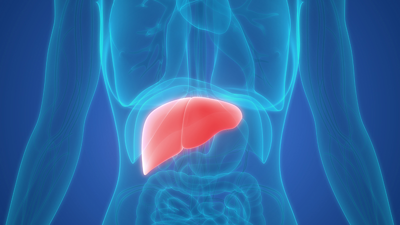
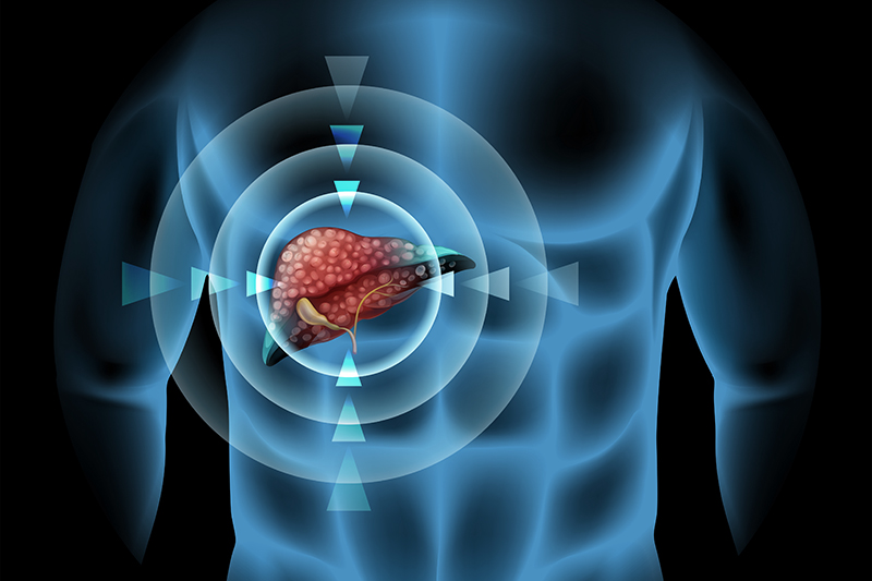
Liver has a complex role in the function of the body, including detoxification, metabolism, bile production, regulation of blood level of sugar, and conversion of excess glucose and fat for storage. Liver cancer is a type of cancer that begins in the cells of your liver. Primary liver cancer (hepatocellular carcinoma) tends to occur in livers damaged by chronic infection, alcohol abuse, and cirrhosis. Cancer starts when abnormal liver cells begin to grow out of control. Most people do not have signs and symptoms in the early stages of primary liver cancer.
Causes
- Cirrhosis due to chronic hepatitis B and C infection
- Alcohol abuse
- Fatty liver
- Diabetes
- Obesity
- Exposure to aflatoxins – found in contaminated peanuts and dried chillies

Signs and symptoms
- Upper abdominal pain
- An enlarged liver, felt as a mass under the ribs on the right side
- Loss of appetite, unintentional weight loss
- Yellowing of the skin and eyes (jaundice)
- Nausea, vomiting
Diagnosis
Tests and procedures used to diagnose liver cancer includes as follows:
- Ultrasound – is often the first imaging used. Additional imaging will be required if any abnormality is found.
- Serum tumor marker – Alpha-fetoprotein blood test (AFP) is used to screen for liver cancer. If AFP levels are very high in someone with a liver tumor, it can be a sign that liver cancer is present.
- CT scan with contrast – produces detailed cross-sectional images of your body. This imaging technique is not suitable for patients with renal impairment and should not be done often.
- Magnetic resonance imaging (MRI) – provides detailed images of soft tissues in the body. MRI scans can be very helpful in looking at liver tumors. Sometimes they can tell a benign tumor from a malignant one. This imaging technique cannot be done in patients with a metal implant.
- Liver biopsy – The doctor will insert a thin needle through your skin and into your liver to obtain a tissue sample. In the lab, doctors examine the tissue under a microscope to look for cancer cells.

Treatment
Treatments for primary liver cancer depend on the stage of the disease. The treatment options include:
- Surgery – In certain situations, your doctor may recommend an operation to remove the liver cancer. However, a small portion of healthy liver tissue that surrounds the tumor also needs to be removed. Therefore, the patient must have good liver function in order to undergo the surgery. Only 10-20% of patients with liver cancer can be treated with surgery.
- Radiofrequency ablation (RFA) – Electric current is used to heat and destroy cancer cells by using an ultrasound or CT scan as a guide. This is a common treatment method for small tumors.
- Transarterial Chemoembolization (TACE) – uses a catheter to deliver both chemotherapy medication and embolization materials into the blood vessels that lead to the tumor. This allows doctors to treat tumors that cannot be treated by surgery. It is a common treatment method for 7-10 cm tumor. Some patients may be asked to return for further treatment, depending on the size, number and location of the tumors.
- Microwave ablation – is quite similar to radiofrequency ablation. In this procedure, microwaves transmitted through the probe are used to heat and destroy the abnormal tissue. The tumor has to be less than 5 centimeters. This procedure can be done using ultrasound or CT-scan as a guide.

Fusion ultrasound – improving outcomes in liver cancer treatment
Treatments for liver cancer depend on the stage of the disease as well as your age, overall health and personal preferences. Operations used to treat liver cancer include: open surgery to remove the tumor and minimally invasive local therapy. In minimally invasive local therapy, an interventional radiologist uses the advanced visualization technology in order to determine the lesion and perform local ablation. This reduces hospital stay and complications. This new imaging technique allows the doctor to conduct the procedure with ease and confidence.
With technological advancement, there is a new imaging technique, called “fusion imaging”. This allows the doctor to fuse CT-scan/MRI images with live ultrasound so they can make confident decisions even in challenging diagnostic cases. Real-time fusion imaging can simultaneously fuse two different images and result in 4D image with increased accuracy. This allows the doctor to see internal organs and tumor clearly.
This new fusion imaging allows the doctor to see the tumor in the liver clearly and precisely. This can increase monitoring and targeting confidence during the procedure. Patients will receive an effective treatment that has low rate of complications and less recovery time.
Catching cancer early often allows for more treatment options. It is often hard to find liver cancer early because signs and symptoms often do not appear until it is in its later stages. Having an annual physical check-up is important in order to screen for liver cancer and identify the risks such as hepatitis b and c infection. Also, eating healthy diet, regular exercise, and avoiding alcohol drinking can help reduce the risks of liver cancer.